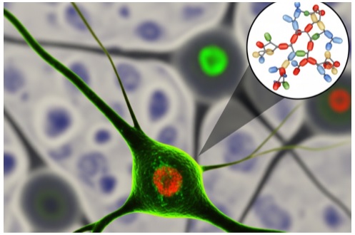
Neuroscience is the study of the nervous system. This includes the brain, spinal cord, and nerves. It controls everything from our heartbeat and breathing to our thoughts, emotions, and actions.
Neuroscience research helps scientists study how the brain works and treat various brain disorders, including Alzheimer’s, Parkinson’s, Epilepsy, and Depression. It also helps doctors find better ways to help people who have had brain injuries or strokes.
When a person suffers from a disease, changes happen inside the brain – these changes can range from harmful protein buildup and inflammation to loss of brain cells and nerve damage. Scientists need to understand these changes in order to develop drugs and treat related diseases. Here is where the Ig secondary antibody comes into play.
What Are Ig Secondary Antibodies?
Ig secondary antibodies are lab-made proteins used in research. They don’t attach directly to the proteins inside the brain. Instead, they bind to primary antibodies, which are already connected to the proteins that scientists want to study.
These secondary antibodies often have special labels on them — like enzymes, color dyes, or fluorescent tags — that help scientists see or measure the proteins. This makes them easier to detect under a microscope or in other lab tests.
What is the Role of Ig Secondary Antibodies in Neuroscience Research?
1. See Brain Cells and Structures
Scientists use techniques like immunofluorescence and immunohistochemistry to study brain tissue. These methods use antibodies to find specific proteins in neurons and other brain cells. Ig secondary antibodies with fluorescent tags help light up these proteins. As a result, researchers can see where they are in the brain.
For instance:
To identify neurons in brain tissue, a scientist may use a mouse antibody that sticks to a neuron protein (NeuN). Then, a goat anti-mouse IgG secondary antibody tagged with a green fluorescent dye (like Alexa Fluor 488) is added. As a result, all the neurons glow green under the microscope. This helps the scientist study their shape and location.
2. Detect Brain Disease Markers
Some brain diseases lead to the buildup of harmful proteins. Scientists use immunohistochemistry and immunofluorescence to find these proteins in brain samples. These methods use primary antibodies to bind to the disease markers and labeled Ig secondary antibodies to make it visible.
For instance:
In Alzheimer’s disease, a harmful protein, known as amyloid-beta, builds up. This leads to the formation of sticky clumps and plaques between brain cells.
Scientists use a rabbit anti-amyloid-beta primary antibody that sticks to the amyloid-beta protein. After this, they add an enzyme-labeled goat anti-rabbit IgG secondary antibody to identify the location of the protein. When a secondary antibody is added, it leads to brown spots on the protein and helps scientists find the presence of amyloid-beta.
3. Measure Protein Levels
Sometimes, scientists not only need to identify the presence of protein. Instead, they need to determine the amount of protein in a sample. At times, they use enzyme-linked secondary antibodies in techniques like WB and ELISA. These antibodies make these proteins show up as a signal. The higher the signal, the higher the amount of protein.
For instance:
A researcher is studying tau protein in Alzheimer’s disease. They run a Western blot using a mouse anti-tau primary antibody. Then they add an HRP-linked goat anti-mouse IgG secondary antibody.
When a detection chemical is added, the amount of light or color produced tells how much tau protein is in the sample. After this, the signal is compared to the healthy brain sample. The difference between the signals indicated how much the disease progresses.
4. Study Multiple Proteins at Once
The brain is complex. Sometimes, researchers need to look at more than one protein in the same sample. They use different Ig secondary antibodies with different colored tags. This allows them to study how various proteins interact or where different cell types are located in the brain. It helps build a more complete picture of what is happening inside the nervous system.
For instance:
To study how neurons and astrocytes (a type of support cell) interact, a researcher uses two primary antibodies:
- Rabbit anti-GFAP to mark astrocytes
- Mouse anti-NeuN to mark neurons
Then, they use:
- Donkey anti-rabbit IgG Alexa Fluor 594 (red)
- Donkey anti-mouse IgG Alexa Fluor 488 (green)
Under the microscope, neurons glow green, astrocytes glow red, and the scientist can see their locations and interactions in the same brain section.
5. Sort Brain Cells
Sometimes scientists want to study just one type of brain cell in detail. To do this, they separate cells using a method called flow cytometry.
Cells are first tagged with primary antibodies that bind to specific surface proteins. Then, Ig secondary antibodies with fluorescent dyes are added to make those tagged cells glow. The glowing cells can then be separated and studied.
For instance:
To study microglia (the brain’s immune cells), a researcher might use a rat anti-CD11b primary antibody to bind to a protein found on microglia. Then, they add a fluorescent goat anti-rat IgG secondary antibody. When the sample is run through a flow cytometer, the glowing microglia are separated from other cells and can be analyzed or grown in culture.
The Bottom Line
Ig secondary antibodies are essential tools in neuroscience research. They make it easier to visualize brain structures, detect disease markers, measure protein levels, study multiple proteins at once, and sort specific brain cells.
These applications help scientists better understand how the brain works and what goes wrong in conditions like Alzheimer’s, Parkinson’s, and other neurological diseases. With this knowledge, they can work toward more accurate diagnoses, effective treatments, and eventually, cures.By lighting the path — quite literally — Ig secondary antibodies help bring clarity to the complex world of the human brain. However, before conducting an experiment, make sure you purchase secondary antibodies online from a reliable source. Otherwise, you may end up having wrong and unreliable results, which can further affect your study.


